how do they x ray babies hips
When A Young Baby Has A Fever It Can Mean That There Is A Serious. This image shows the soft.

Hip Dysplasia Should My Child Be Screened Uva Radiology
How do they x ray babies uk are a topic that is being searched for and liked by netizens today.

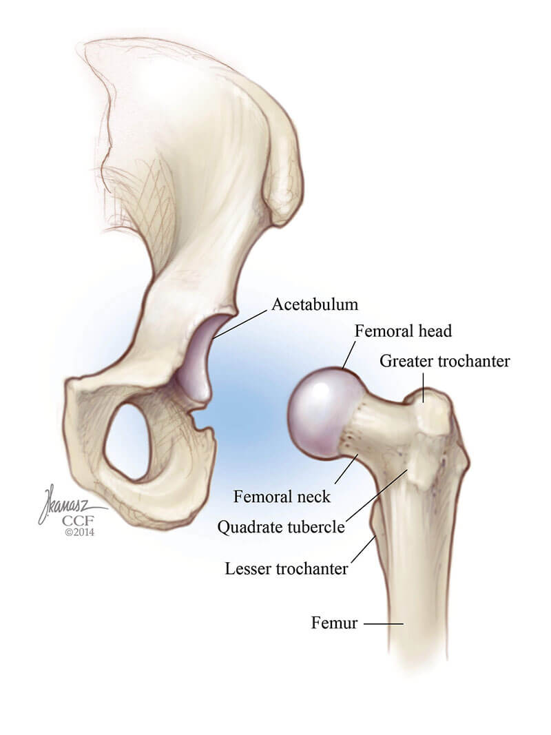
. It s sometimes called congenital. The doctor hears or feels a hip click when moving the infants thigh outward during a routine checkup. An X-ray of the pelvis focuses specifically on the area between your hips that holds many of your reproductive and digestive organs.
Youll be asked to. If she does have it they may try to brace it first. A hip X-ray radiograph is a medical imaging test that creates a.
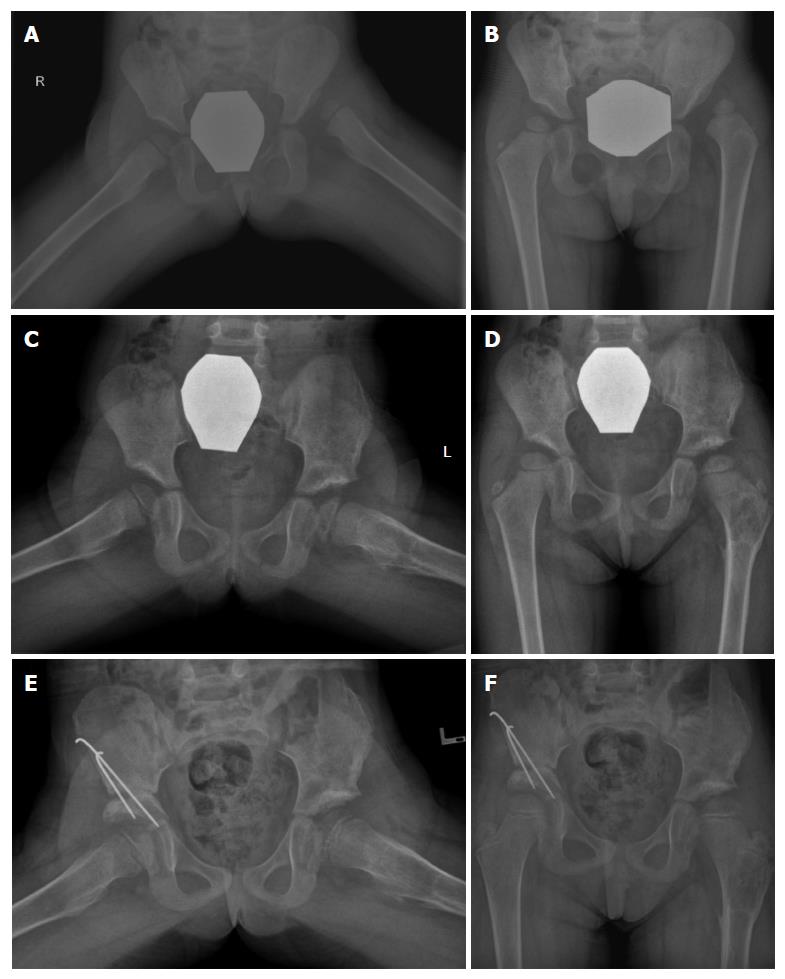
For children with normal physical exams referral is warranted for any child with dysplasia or shallowness of the hip joint beyond 6 weeks of age. You will go in the room with him he will need to be stripped from the waist down they will take x-rays of him flat on his back legs dead straight. In these younger children it is possible to nip the problem early with night time bracing to help the hips recover if hip dysplasia returns.
The babys legs have differences in their lengths or appearances. Lying on his back one leg in the knee bends at an angle. The main pathology is.
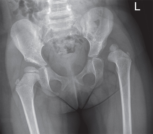
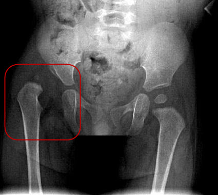
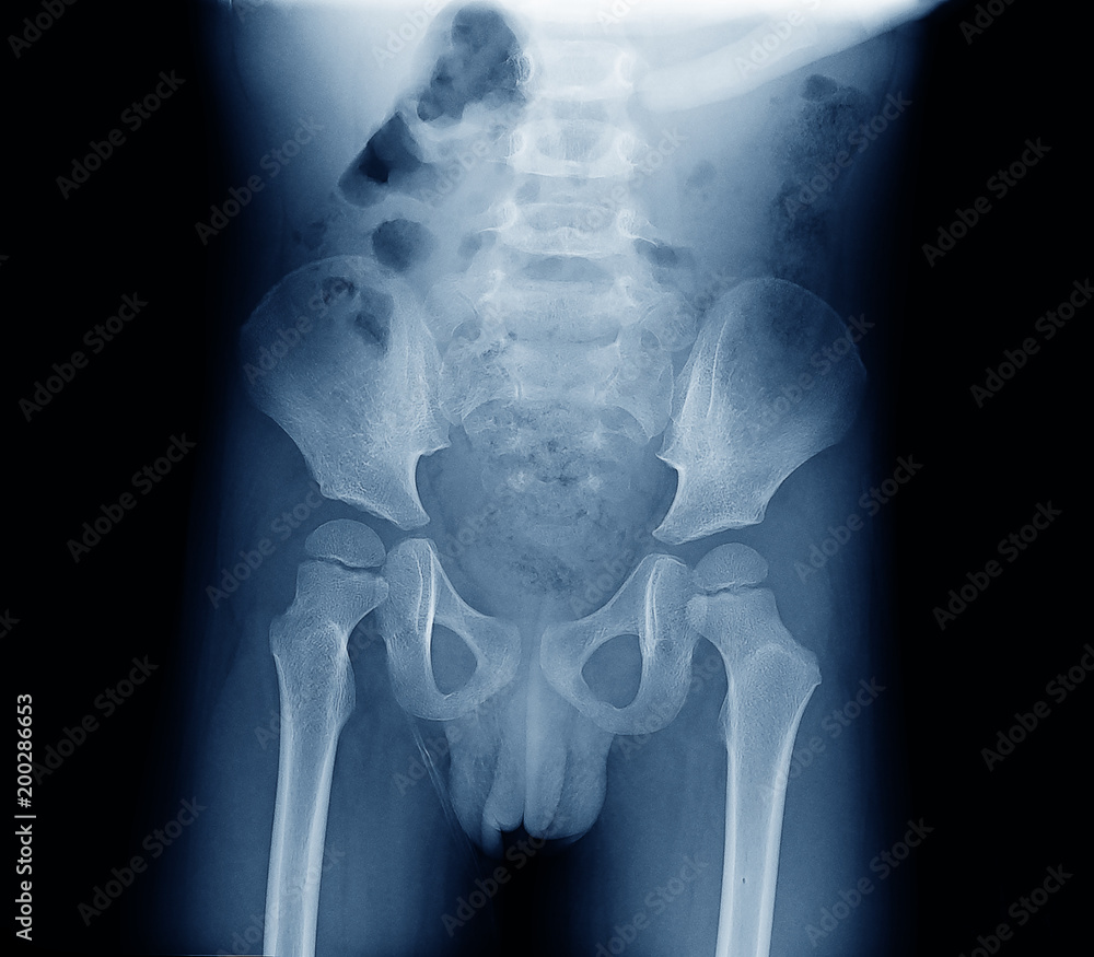
If an X-ray of the hip joints is performed according to Launstein Lauenstein then the patients position looks like this. How do they x ray babies hips. In a hip X-ray an X-ray machine sends a beam of radiation through the pelvic bones and hip joints where the legs attach to the pelvis and an.
Its a cast that goes around both hips and down the leg to keep the hips. Your How do they x ray babies uk images are available. Totaleclipse 07092007 1731.
A hip X-ray is a safe and painless test that uses a small amount of radiation to make images of the hip joints where the legs attach to the pelvis. Developmental dysplasia of the hip. An ultrasound machine sends sound waves into the hip area and images are recorded on a computer.
But any time you are. The authors of the second paper also evaluated the. Two tests are performed called the Barlow.
During the examination an X-ray machine sends a beam of radiation through the pelvic bones and hip joints and an image is recorded on a computer or special film. If it persists they may put on a spica cast.
Developmental Dysplasia Of The Hip Radiology Reference Article Radiopaedia Org

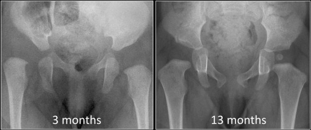
Acetabular Remodeling After Closed Reduction Of Developmental Dysplasia Of The Hip

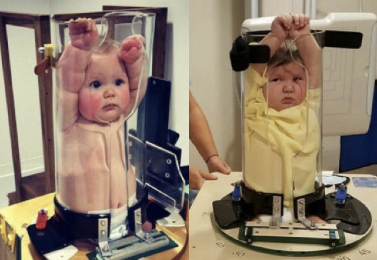
Ce4rt Guide For X Ray Techs To Immobilize Pediatrict Patients
Hip Dysplasia Symptoms Causes Treatments Tests Recovery

What Pregnancy And Childbirth Do To The Bodies Of Young Girls The New York Times
Adolescent Hip Dysplasia Orthoinfo Aaos

X Ray Exam Pelvis Connecticut Children S

Infant Diagnosis International Hip Dysplasia Institute

Diagnosis Of Hip Dysplasia Healthy Hips Australiahealthy Hips Australia
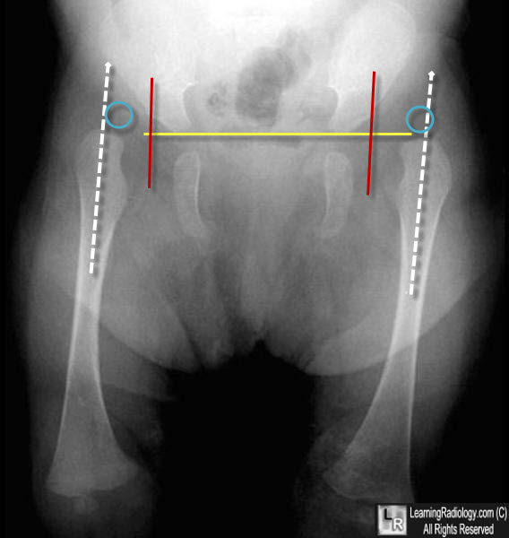
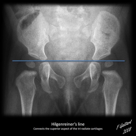
Learningradiology Developmental Dislocation Dysplasia Of The Hip

Sample Image Of Baby Hip X Ray With Image Quality Review Areas Highlighted Download Scientific Diagram

Hip I Developmental Dysplasia Of The Hip Musculoskeletal Key
Developmental Dysplasia Of The Hip Prof Portinaro Orthopedic Suregon
Developmental Dysplasia Of The Hip What Has Changed In The Last 20 Years
Baby X Rays Look Like This And Twitter Is Making Hilariously Dark Jokes
Modern Approach To Developmental Dysplasia Of The Hip Mayo Clinic
The Radiology Assistant Developmental Dysplasia Of The Hip
X Ray Screening International Hip Dysplasia Institute
X Ray Image Of Child Bone Show Pelvis Hip Joint Spine Stock Photo Adobe Stock